The University of Oregon does not comment on injuries, leaving many left speculating as to how severe an injury may actually be. Football is a rough sport, one that causes physical harm to those who play, and inevitably bones break and muscles and tendons tear.
Such has been the case early on in the 2012 football season for the Oregon Ducks–through two games already Oregon Ducks athletes have suffered major injuries that will end their seasons. Some injuries are still unofficial, but two major losses have been made official.
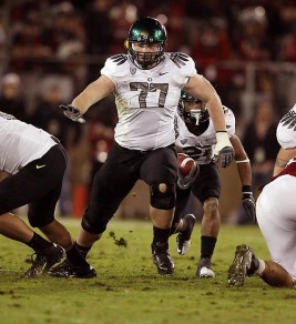
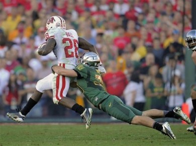
Senior Safety All-American Candidate John Boyett’s year is done, he is having surgery on both knees to repair tears in his Patellar Tendons.
Senior Guard Carson York is also done for the year, having broken his knee cap in the game against Fresno State.
Both are huge losses for Oregon, both in on-field play and leadership. Barring medical redshirts granted, fans have unfortunately seen the last of two of the Ducks most important players in an Oregon uniform.
The news of both came as quite a shock, but what does it mean? What happens when a Patellar Tendon tears, and why will Boyett miss the entire season because of it? And how exactly does somebody break a kneecap, and what is needed to repair it?
In this article, we’ll take a look at both injuries, the procedures needed to fix it, and the expected recovery time for both student-athletes.
Patellar Tendon Tears
(John Boyett’s Injury)

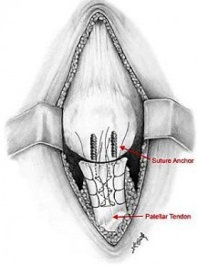
- Figure 1: Knee Showing Patellar Tendon Tear
John Boyett is scheduled to have surgery on both knees to repair partial tears of both Patellar Tendons. These injuries did not occur during the game, they have been lingering injuries developing worse over time.
Boyett could have, and perhaps should have, had surgery to repair the issue last year or during the off-season, but seeking out specialist advice there was thought that perhaps he could play through the injuries, strengthening his knees elsewhere to compensate (reported upon announcement of his injuries).
Partial tears: Many tears do not completely disrupt the soft tissue. This is similar to a rope stretched so far that some of the fibers are torn, but the rope is still in one piece.
Complete tears: A complete tear will disrupt the soft tissue into two pieces.
The patellar tendon often tears where it attaches to the kneecap, and can break a piece of the bone as it tears. When the patellar tendon is completely torn, the tendon is separated from the kneecap. Without this attachment, the knee cannot be straightened.
Injury
A very strong force is required to tear the patellar tendon.
Falls. Direct impact to the front of the knee from a fall or other blow is a common cause of tears. Cuts are often associated with this type of injury.
Jumping. The patellar tendon usually tears when the knee is bent and the foot planted, like when landing from a jump or jumping up.
Tendon Weakness
A weakened patellar tendon is more likely to tear. Several things can lead to tendon weakness.
Patellar Tendonitis
Inflammation of the patellar tendon, called patellar tendonitis, weakens the tendon. It may also cause small tears. Patellar tendonitis is most common in people who participate in activities that require running or jumping. While it is more common in runners, it is sometimes referred to as “jumper’s knee.”
Corticosteroid injections to treat patellar tendonitis are typically avoided in or around the infrapatellar tendon. Injections around this articular tendon have been linked to increased tendon weakness and increased likelihood of tendon rupture.
Surgical Treatment
Most people require surgery to regain the most function in their leg. Surgical repair reattaches the torn tendon to the kneecap.
People who require surgery do better if the repair is performed early after the injury. Early repair may prevent the tendon from scarring and tightening in a shortened position.
Hospital stay
Tendon repairs are sometimes done on an outpatient basis. Most people do stay in the hospital at least one night after this operation. Whether or not an overnight stay is needed will depend on medical needs.
The surgery may be performed with regional (spinal) anesthetic, or with a general anesthetic (breathing tube). It cannot be done under local anesthesia.
Procedure
To reattach the tendon, sutures are placed in the tendon and then threaded through drill holes in the kneecap. The sutures are tied at the top of the kneecap. The surgeon will carefully tie the sutures to get the correct tension in the tendon. This will also ensure the position of the kneecap closely matches that of the uninjured kneecap.
Rehabilitation
After surgery the patient will require some type of pain management, including ice and medications. About two weeks after surgery, skin sutures or staples will be removed in the surgeon’s office.
 Most likely, the repair will be protected with a knee immobilizer or a long leg cast. The patient may be allowed to put weight on the leg with the use of a brace and crutches (or a walker).
To start, the surgeon may recommend “toe touch” weight-bearing. This is when the patient lightly touches their toe to the floor, putting down just the weight of the leg. By two-to-four weeks after surgery, the leg can usually bear about 50% of body weight. After four-to-six weeks, the leg should be able to handle full body weight.
Over time, the doctor or therapist will unlock the brace. This will allow the patient to move more freely with a greater range of motion. Strengthening exercises will be added to the rehabilitation plan.
In some cases, an “immediate motion” protocol (treatment plan) is prescribed. This is a more aggressive approach and not appropriate for all patients. Most surgeons protect motion early on after surgery.
The exact timeline for physical therapy and the type of exercises prescribed will be individualized. Each patient’s rehabilitation plan will be based on the type of tear, surgical repair, medical condition, and the patient’s needs.
Complete recovery takes about six months. Many patients have reported that they required 12 months before they reached all their goals.
It is sad to see John Boyett’s career end this way after such an impressive three-year stretch. Had Boyett had this surgery during the off-season, he may have been able to play this year, but recovery times vary widely. Here is wishing for John’s recovery to be complete and that he can resume his full life after this is all done, be it continuing to play football at the next level or pursuing a career elsewhere.
Broken Patella (kneecap)
(Carson York’s Injury)
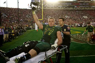
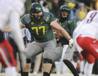
Still not at 100% in recovery from his knee injury in the Rose Bowl, during the game vs. Fresno State last Saturday Carson York suffered a Broken Patella, ending his participation in the 2012 season. As a senior, it likely means his career at the University of Oregon is over.
A patella fracture is an injury to the kneecap. The kneecap is one of three bones that make up the knee joint. The patella is coated with cartilage on its under-surface, and is important in providing strength of extension (straightening) of the knee joint.
 Causes of a Patella Fracture
A patella fracture most often occurs from a fall onto the kneecap. When the fracture occurs due to this type of direct trauma, there is often damage to the overlying skin, and because of the limited amount of soft tissue this can easily become an open fracture.
Patella fractures can also occur when the quadriceps muscle is contracting but the knee joint is straightening (a so-called “eccentric contraction”). When the muscle pulls forcefully in this manner, the patella can fracture.
Patella Fracture Treatment
Patella fractures should be seen in the emergency room. X-rays will determine the type of fracture and the amount of displacement (separation) of the fracture. One of the critical factors in determining treatment is a thorough examination–specifically, doctors will check if the patient can perform a straight-leg raise.
Diagnosis
A straight-leg raise test is done by having the patient lie flat on a bed. With the leg straight, the patient should then raise his or her foot off the bed and hold it in the air. This tests the function of the quadriceps muscle and its attachment to the shin bone (Tibia). A disruption of the quadriceps tendon, patella or patellar tendon can lead to inability to perform a straight-leg raise. If a straight-leg raise can be done, then non-operative treatment may be possible in the setting of a patella fracture.
One of the common symptoms of a patella fracture is knee swelling. The swelling is caused by bleeding from the fractured bone ends into the knee joint. Patients with a large amount of blood in the knee may benefit from draining the blood for pain relief. Immobilizing the knee with a knee brace will also help minimize discomfort.
Patella Fracture Surgery
Patients with non-displaced (not separated) or minimally displaced fractures who can perform a straight-leg raise (as described above) can usually be treated without surgery. A long leg cast or a knee immobilizer can be used for treatment of these types of patellar fractures.
When surgery is necessary, an incision is made over the front of the knee joint. The fractured ends of bone are realigned and held in place with some combination of pins, screws and wires. In some cases, a portion of the patella can simply be removed, but this is usually done for smaller fracture fragments.
Rehab after Surgery
Following surgery, patients will need to keep their knee in a straight position to allow for initial healing. Exactly when the knee can begin moving depends on the strength of the repair the surgeon is able to achieve. Gentle motion can usually begin in the first weeks following surgery. In situations where the bones are held solidly, early motion of the knee helps to achieve the best results after surgery.
The most common complication of patella fracture surgery is that the metal implants can be painful over time–especially when kneeling. Because of this, it is not uncommon for a second procedure to remove the metal implants. This procedure is usually done at least a year after the initial surgery.
Other possible complications include:
Infection
Non-healing fractures
Failure of the fixation to hold the fragments in place
Kneecap pain (chondromalacia)
Knee arthritis
Like with John Boyett’s injury, my feelings go out to Carson. We hope he gets well soon and can return to the life he want as soon as possible!
and with that, the doctor is out.
Related Articles:
NeuroDocDuck (Dr. Driesen) is a doctor who specializes in neurology, and sports medicine. He is an Oregon alumnus, completing his medical education and training in the UK. He has been both a practicing clinician and professor, a well-known and respected diagnostician, an author, and has appeared on national television.
NeuroDocDuck is active in his profession, and stays current on all new trends in his field. He enjoys golf and loves his Ducks!


